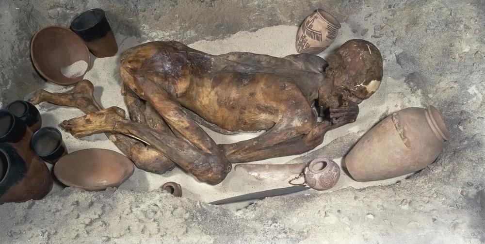
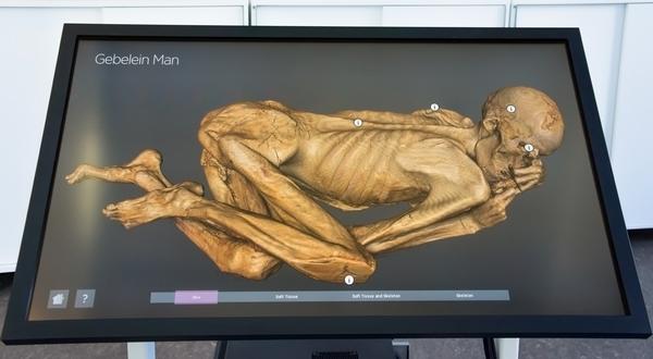
In November 2012, curators at the British Museum in London, UK, collaborated with a team of medical experts to create a computerised tomography (CT)-scanned mummy. From the scans, new insights were gained into life and death in Ancient Egypt more than 5,000 years ago.
The same technology has potential for use in the attractions industry, not least because it offers an exciting new way for visitors to experience data.
The groundbreaking techniques, which digitally unwrapped the mummy layer by layer, revealed unprecedented information about his age, health and even diet, as the technology uncovered hidden interior details that had not been seen before.
The data gathered was transformed into 3D visualisations thanks to volume graphics software like that used in the auto engineering industry.
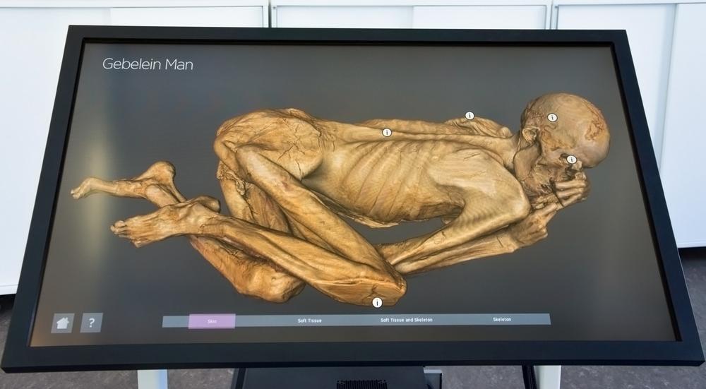
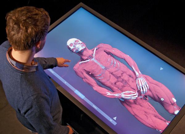
A temporary exhibition, Virtual Autopsy: Exploring a Natural Mummy from Early Egypt, at the British Museum, used detailed images created from high-resolution CT scans to build interactive installations. These 3D digital installations, using visualisation software known as Inside Explorer, allowed visitors to delve into the interior layers of the mummy, thanks to the zooming, panning, rotating and peeling functions available via an interactive touch screen.
The success of Virtual Autopsy led to the installation of a permanent exhibition at the British Museum and a second temporary exhibition last year entitled Ancient Lives, New Discoveries. The exhibit presented the scans of eight mummified humans from various points in history, using pre-rendered video sequences.
TECHNOLOGICAL ADVANCES
The technology is improving. It’s becoming easier to use and producing ever more comprehensive results. In fact, the technology is the same as that used in hospitals for clinical work and for human forensic investigation.
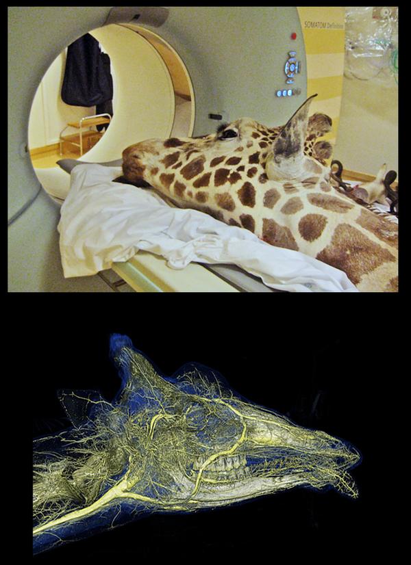
Owner of the software, the Interactive Institute Swedish ICT, has identified interest from zoos and aquariums in using the same technology to offer visitors a new perspective on the creatures in front of them. As the cost and radiation dosages of modern CT scanners come down and image quality improves, CT scanners are starting to be used more frequently in zoos and veterinary clinics.
“The technology is getting better images with lower dosages, which has always been one of the goals of scanner manufacturers,” says David Hughes, manager of solution development at Interactive Institute Swedish ICT.
APPLICATIONS IN ZOOS
The scans of the animals, as well as being used for medical purposes, could also potentially be used to engage visitors, not only in science and history museums, but in a wider range of sectors in the attractions industry.
“Public engagement activities around human anatomy in science centres could be replicated in zoos and potentially aquariums,” he says. “There’s no real reason as to why this couldn’t be repeated, certainly with vertebrates.”
The scanning of live animals presents several key points that could be interesting to educators in attractions. The data and its format offers new perspectives to the visitor, the scans can be used to tell stories about the animals being highlighted and to illustrate the work of animal carers and they can facilitate a deeper understanding of what’s in front of the visitor’s eyes.
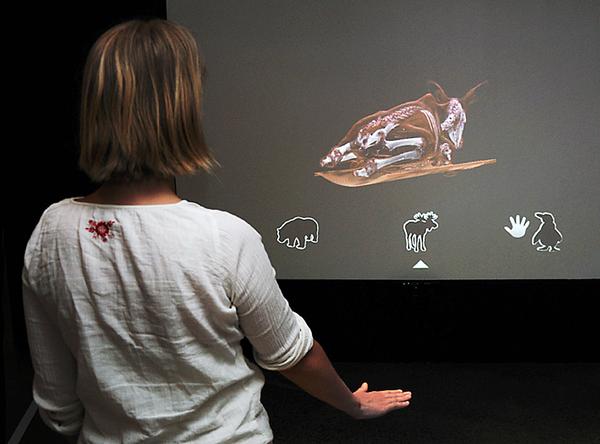
Interactive Institute Swedish ICT worked with Visualisation Centre C, Centre for Medical Image Science and Visualisation (CMIV) and Kolmården Zoo, Sweden, on an exhibition, built around authentic scan images of animals – a moose, a penguin and a brown bear. Visitors used arm and hand gestures to interact with the 3D display. This first-of-its-kind exhibit could be the start of something new for zoos.
CLOSER TO RESEARCH
“This was a part of an exhibition called Animalistic, which described how scientists are using state-of-the-art technology to understand how animals live, behave and perceive the world,” says Hughes. “We’ve received a lot of interest from aquariums and zoos worldwide, who see Inside Explorer as a way of creating new user experiences bringing visitors closer to the research.”
In fact, the success of Inside Explorer has meant the team at the institute has started up a new dedicated company called Interspectral that will continue to develop and market the Inside Explorer solution and provide services in the 3D digitisation domain. This is normal practice for the institute, which takes promising research, incubates solutions around it and spins them out.
“With a sick animal, you could present the latest developments in its recovery through this technology,” Hughes says. “If an animal broke a leg, you could have a public station where visitors would follow the story of the treatment and recovery. That could bring people back regularly and it would promote the work of the veterinary side of an institution.”
“Animals have health challenges, as we do, and I think there are stories to tell there that aren’t being told at the moment. The scans are there so why not use them?” Hughes says.
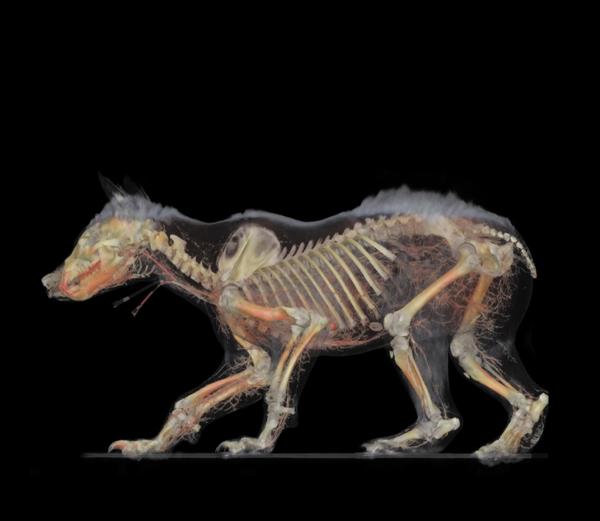
PHYSIOLOGICAL INSIGHTS
“The technology also provides great insights into animal physiology,” he continues. “What does a leopard look like on the inside? Why is it built the way it is? Scans reveal the orientation of the bones and muscles, and visitors can learn how a leopard’s abilities are reflected in its physiology. An installation could show a visitor details inside the animal even while he’s looking at the real thing, so he can understand why they are the way they are. It’s something that’s not really been done and I think it would be fascinating.”